3D Slicer 4.8 gets Released with Improved DICOM support and Segmentation Capabilities


3D Slicer is a free, open source, extensible application for visualization and image analysis. It is widely used in the field of medical computing, with over 325,000 downloads. In the field, 3D Slicer is applied to research and applications that range from preclinical animal studies to surgical planning, surgical guidance, medical robot control, 3D printing and population studies.
On behalf of the 3D Slicer development community, we are pleased to announce that version 4.8 is now available for download. This version introduces close to 1,000 feature enhancements and bug fixes for better performance and stability. It includes more than 15 new-and-improved core modules and dozens of updated extensions.
Here are some release highlights:
- a simpler Data module, which integrates Subject Hierarchy;
- improvements to the Segmentations module that add effects like Grow from seeds, Fill between slices, Surface cut, Mask Volume, Watershed, Fast marching and Flood Filling;
- improvements to slice viewers, including better crosshair usability and support for slice model projection;
- support for Volumetric Meshes;
- improvements to DICOM support that enable the definition of semantic meanings and faster imports;
- improvements to the Transforms module, such as support for interactively updating transforms in the 3D view and support for visualizing displacements of individual points;
- support for fast and interactive plotting through VTK Charts; and
- the transition from the mailing list to the 3D Slicer Forum on Discourse.
Please visit the forum for a complete list of improvements and fixes.
The development of 3D Slicer—including its numerous modules, extensions, datasets, pull requests, patches, issues reports, suggestions—is made possible by users, developers, contributors and commercial partners around the world. This development is funded by various grants and agencies. For more details, please see the 3D Slicer Acknowledgments page.
We are one of the lead maintainers of 3D Slicer and other software solutions such as the Insight Segmentation and Registration Toolkit (ITK), the Visualization Toolkit (VTK) and CMake. To support these solutions, we offer consulting services. Contact us at kitware@kitware.com to learn how we can help integrate the software into your research, products and processes.
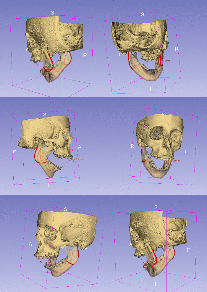
Figure 1: 3-dimensional reconstruction of craniofacial hard tissue (bone, in solid/semitransparent yellow) and soft tissue (vessels in solid red, gingiva in semitransparent pink) structures, rendered altogether with orthodontic appliances (brackets in solid blue) obtained from Cone Beam Computed Tomography. This data was generated as surgical scenario for Bisagittal Split Osteotomy (BSSO) training. The models in this figure represent the pre-surgical state of the patient, and Slicer 4.8 will help the trainee practicing all the different steps of BSSO, in which (1) the gingiva is cut to expose the bone, (2) the bone is cut bilaterally in the mandible into distal and proximal segments, (3) the distal segment of the mandible is moved forward and finally (4) stabilized in its new position in the proximal segment with fixation screws. BSSO is performed to return patient skeletal structures into their proper alignment in cases of mandibular deficiency. This work was supported by the National Institute of Health (NIH) National Institute for Dental and Craniofacial Research (NIDCR) grant R43DE027595 (High-Fidelity Virtual Reality Trainer for Orthognathic Surgery).